Reticular Formation And Associated Diseases
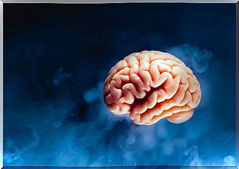
The reticular formation consists of an aggregate of nuclei and nerve fibers that occupy the segment of the brainstem, between the nuclei of the cranial nerves and the ascending and descending nerve pathways.
In some way, all the nuclei of the trunk belong to the reticular formation, with the possible exception of the roots of some cranial nerves; however, it appears that reticular formation is critical for the expansion of the spinal interneuron system.
To date, however, it is not yet fully understood which parts of the nervous system make up the reticular formation. It is believed that its extension starts more or less from :
- The upper part of the spinal cord.
- Brain stem.
- Mid-brain.
- Quadrigeminal tubercle.
- Diencephalon.
- Hypothalamus.
- Subthalamus.
- Thalamus.
The roles of the reticular formation are different. Among them we find:
- Coordination of head and body movements through the stimulation and inhibition of voluntary movements and reflex movements.
- Breathing.
- Blood pressure.
- Sensory response, especially to pain.
- Psychological processes such as arousal or attention.
- Sleeping and dreaming.
- Muscle tone.
A very important part of the reticular formation is the raphe. The frontal nucleus of the raphe projects on some parts of the frontal, parietal and occipital cortices, as well as in cortical areas of the cerebellum related to their functions.
These projections secrete serotonin, a neurotransmitter derived from tryptophan. It therefore seems that the nucleus of the dorsal raphe carries out generalized actions.

The reticular formation
From a physiological point of view, the reticular formation can be seen as a multineuronal postsynaptic formation. It has axons arranged transversely and longitudinally.
It does not transmit particular messages such as sensitive or motor messages. Rather, it receives signals and transmits them to the central nervous system in the form of general information. Depending on the location of the groups of nuclei, the reticular formation consists of:
The central nuclei
They occupy the upper area of the brain stem. They are divided into:
- Ventral nucleus (lower bulb).
- Giant cell nucleus (superior bulb).
- Pontine nuclei.
- Mesencephalic nucleus.
Lateral and paramedian nuclei
The lateral and paramedian nuclei are related to the functions of the cerebellum. They form part of the reticulo-cerebro-reticular circuit, where the fibers of the reticular formation accompanying the spinothalamic bundle project towards the cerebellar cortex.
The fibers composed here, as well as the nuclei in general, intervene on the coordination of reflexes and muscle tone.
Medial nuclei
They consist of:
- Gray substance surrounding the mesencephalic duct.
- The nuclei of the raphe of the bulb.
- The dorsomedial nucleus.
Functional anatomy of the reticular formation
The physiology of reticular formation consists of an aggregate of neurons and axons in charge of associating and combining information from the outside and inside of the nervous system. This system regulates the following:
- Functioning of the nuclei of the cranial nerves.
- Sending of alarm signals to the sensory centers.
- Stimulation of the higher centers, which in response perform an inhibitory or facilitating function on the central nuclei of formation.
- The functions of the cerebellum. Through its projections to the cerebellum it participates in the coordination of reflexes and muscle tone.
- Association between hypothalamus and brainstem.
- Adjust information related to pain perception.
What can happen to the raphe?
According to Arnold E. Eggers, some physiological effects of stress result in increased activity of neurons in the dorsal raphe nucleus.
Stress also produces excitotoxic damage in the hippocampus, which leads to the malfunction of the normal activity of the raphe nucleus. This imbalance can cause or contribute to the cause of a variety of disorders, including:
- Migraine (selective serotonergic hyperactivity).
- Hypertension.
- Cerebrovascular damage.
- Temporal lobe epilepsy (defective neuronal resonators).
- Schizophrenia.
- Post-traumatic stress disorder.
As we have seen, the reticular formation appears to have no limits and is associated with the areas of the brain that we have mentioned. Furthermore, the research carried out so far seems to associate it with some serious brain disorders.
